Comprehensive Fetal Medicine & Advanced Pregnancy Imaging
From Early pregnancy scans to Anomaly scans, Fetal Echocardiography and Genetic testing—we offer world-class Fetal care for every mother and every baby.
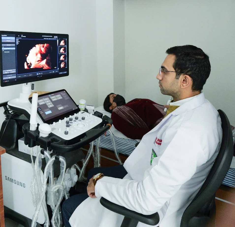
All fetal scans and invasive procedures are personally performed and interpreted by a fellowship-trained Fetal Medicine specialist (Ex-PGIMER Chandigarh) with advanced training in Fetal Echocardiography from Harvard Medical School, Boston (USA)—ensuring the highest standards of diagnostic accuracy and ethical, compassionate care.
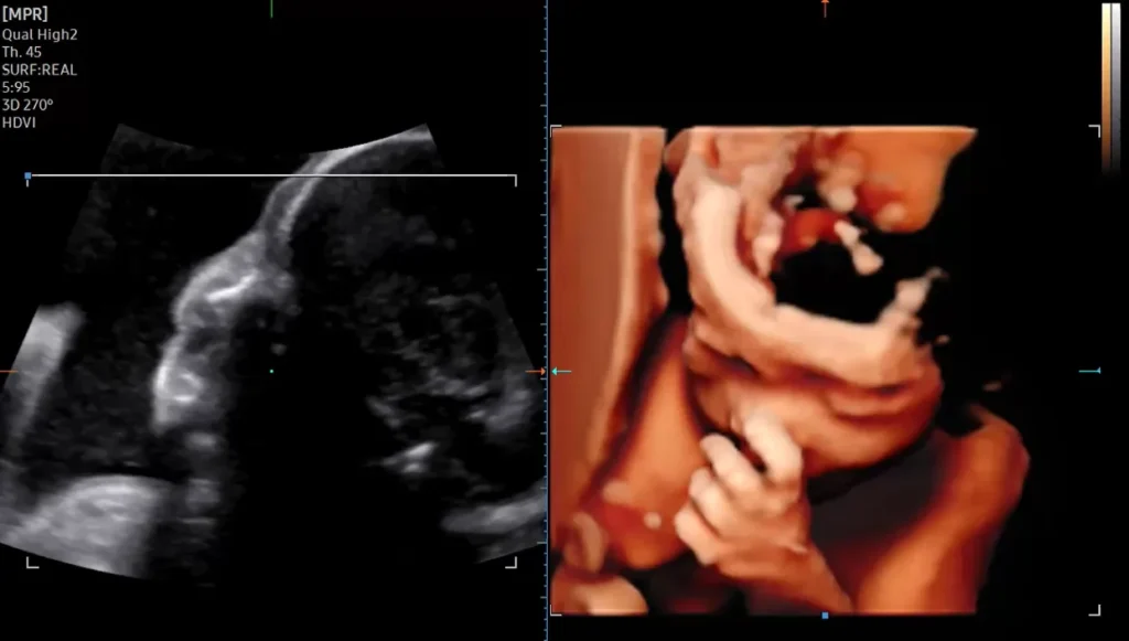
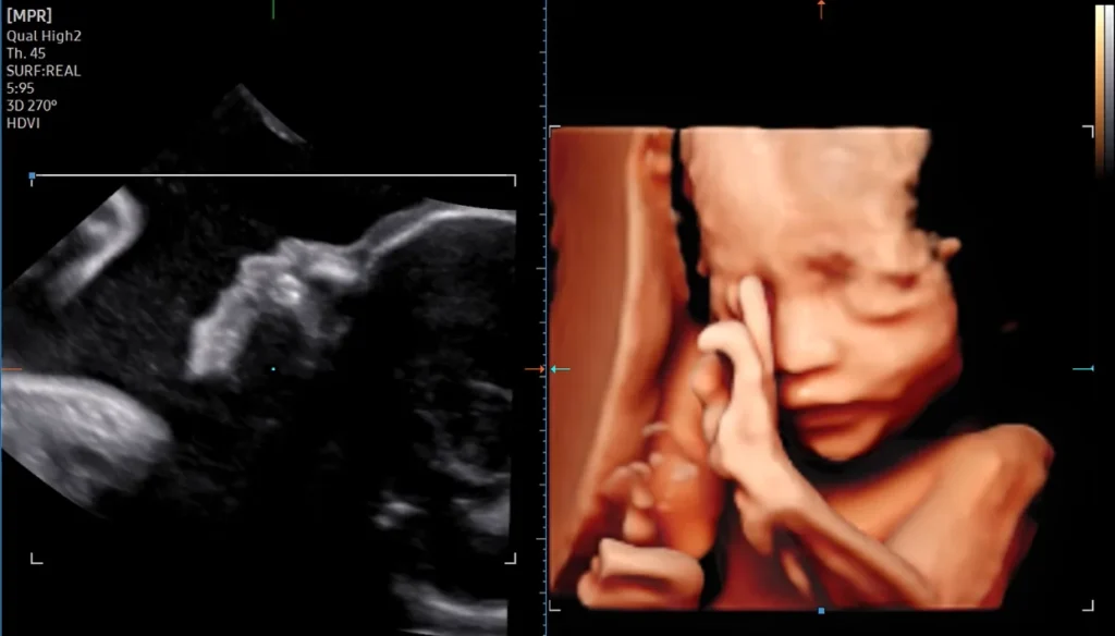
High-Resolution Ultrasound Systems
We use premium-grade ultrasound platforms offering:
Advanced, detailed evaluation of fetal cardiac anatomy and rhythm, performed by a specialist with international training in Advanced Fetal Echocardiography (Harvard, Boston, USA).
Recommended in:
IVF pregnancies
Diabetes
Family history of congenital heart disease
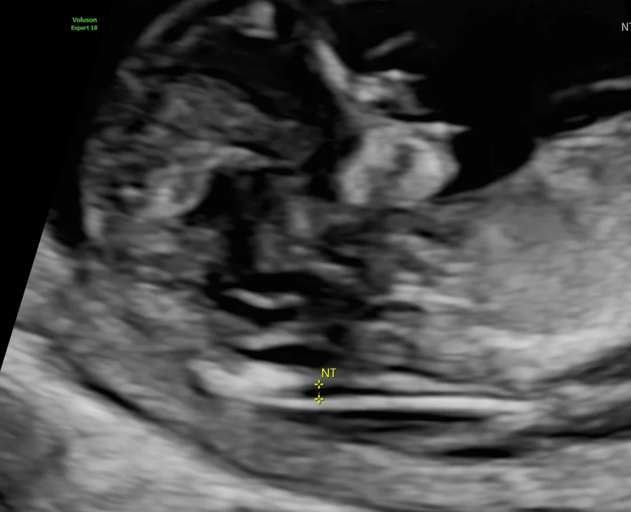
Abnormal NT
Suspected cardiac anomalies
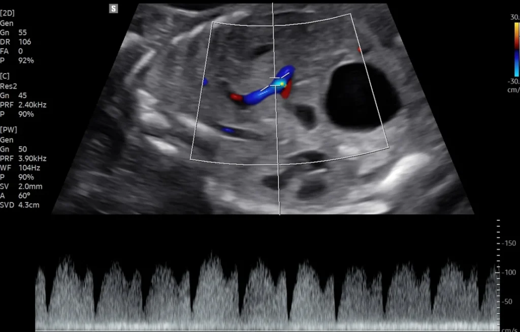
Fetal Doppler Studies (30-34 weeks)
Assessment of fetal circulation in:
IUGR
High-risk pregnancy
Preeclampsia
Twin–Twin Transfusion Syndrome
Gestational diabetes & hypertension
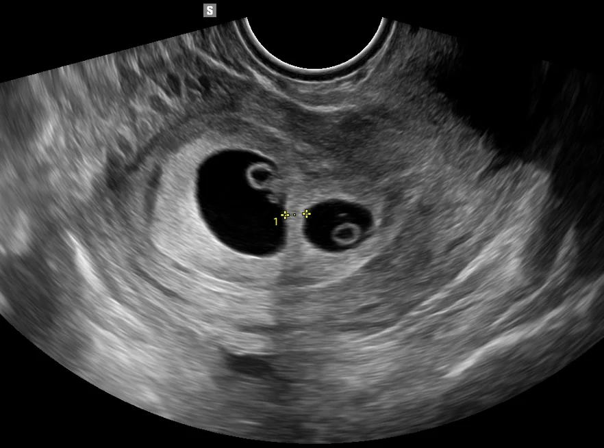
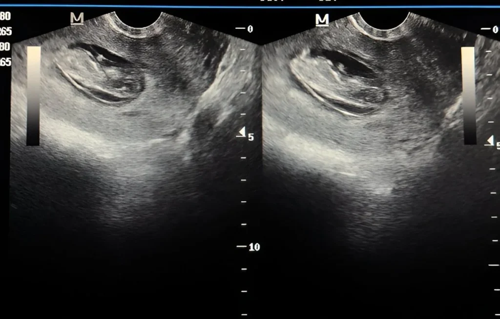
Transvaginal Sonography (TVS)
High-resolution imaging for early pregnancy, placental location, cervix length, previa, and targeted evaluations.
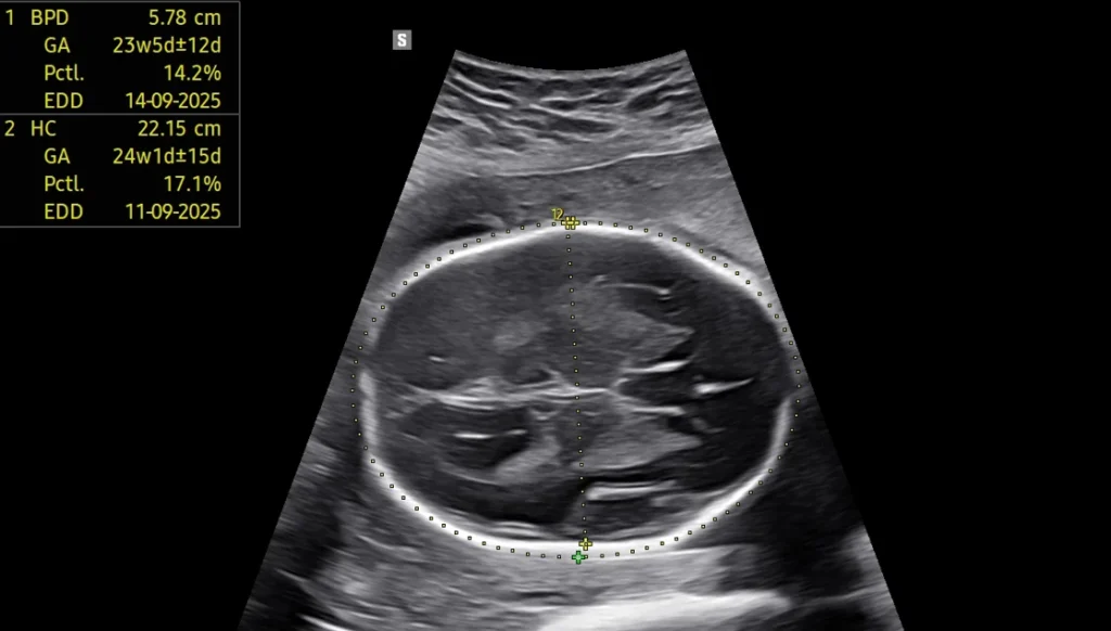
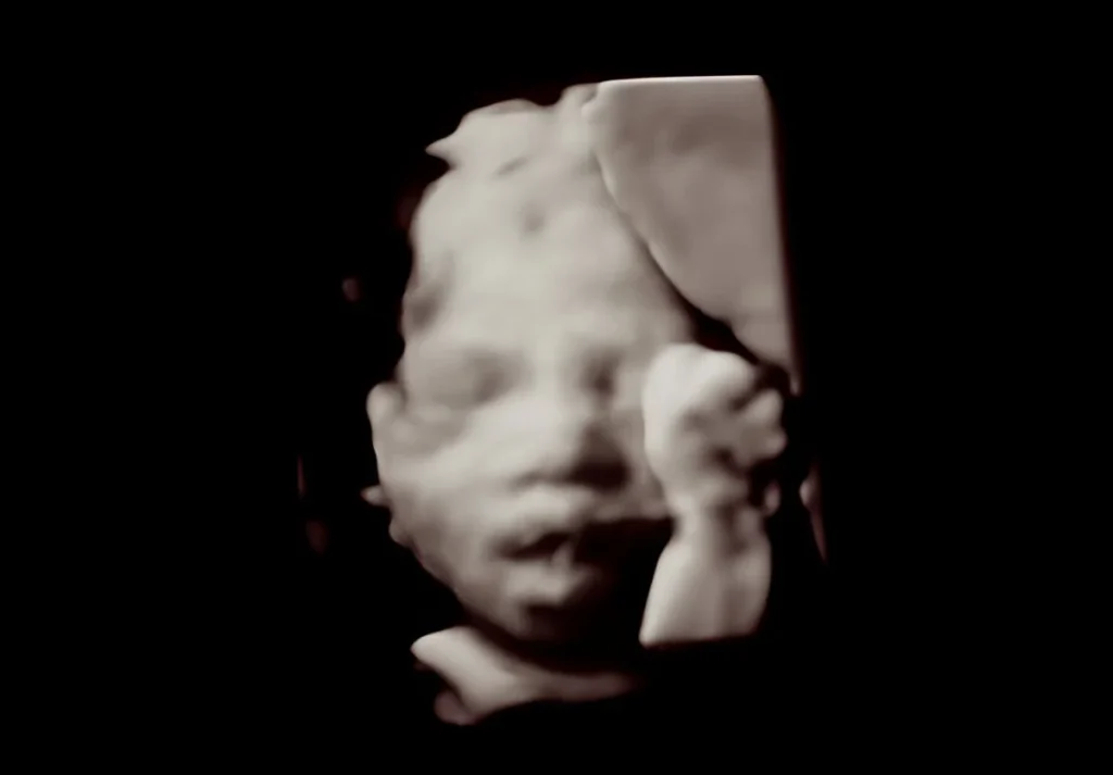
Fetal Well-Being & Biometry
Growth scans, BPP, AFI, interval growth patterns, and third-trimester monitoring.
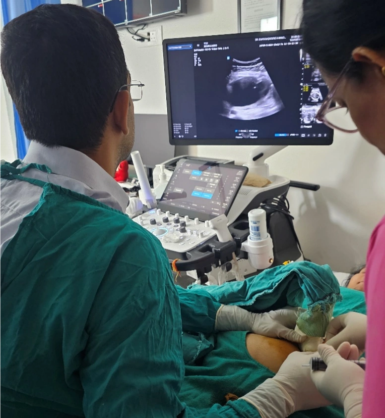
Fetal Interventions
Amniocentesis
Genetic testing & infection workup.
Performed under continuous ultrasound guidance
Minimal-risk technique
Detailed counselling before & after the procedure
Chorionic Villus Sampling (CVS)
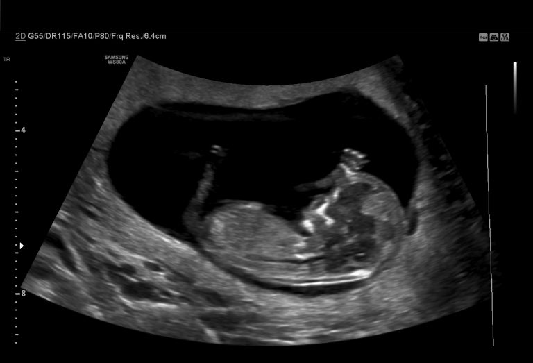
Early genetic diagnosis at 11–13 weeks.
Genetic Services
Dual Marker Test (11–14 weeks)
Combined screening for trisomy risk when done with NT scan.
Quadruple Marker Test (15–22 weeks)
Second-trimester maternal serum screening.
NIPT (Non-Invasive Prenatal Testing)
Highly accurate blood test for trisomy 21, 18, 13 and sex chromosome anomalies.
FISH & Karyotyping
Chromosomal analysis for genetic conditions.
Chromosomal Microarray (CMA)
Advanced genetic evaluation for subtle chromosomal abnormalities.
Whole Exome Sequencing (WES)
Molecular-level genetic diagnosis for complex or recurrent anomalies.
Antenatal Genetic Counselling
Counselling for high-risk results, abnormal scans, family history and recurrence risk.
Preconception Genetic Counselling
For couples planning pregnancy with known risks.
Frequently Asked Questions
Is fetal ultrasound safe?
Yes. Ultrasound uses sound waves and has no radiation.
Do I need a full bladder?
Required only in early pregnancy scans.
How long does a Level II scan take?
Usually 20–30 minutes, depending on fetal position.
When is a Fetal echo needed?
Between 18–24 weeks or earlier if recommended.
Cart
No products in the cart.
Select the fields to be shown. Others will be hidden. Drag and drop to rearrange the order.